RADIOLOGY
RADIOLOGY
Radiology is an all-important medical specialisation that uses imaging techniques to diagnose and treat diseases especially related to internal organs and not externally visible. It encompasses both diagnostic radiology and interventional radiology.
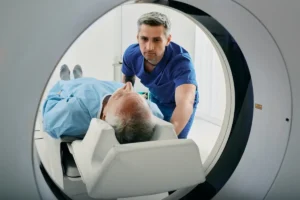
Diagnostic Radiology:
Diagnostic radiology involves using various imaging procedures like X-rays, CT scans, MRI scans, ultrasound, and nuclear medicine scans to visualize the body’s internal structures and identify diseases.
Interventional Radiology:
Interventional Radiology uses imaging guidance (like X-ray fluoroscopy, CT, ultrasound, or MRI) to perform minimally invasive procedures, such as biopsies, angioplasty, stent placement, and catheter insertions.
The various subspecialties of radiology, specific to the areas of the body are:
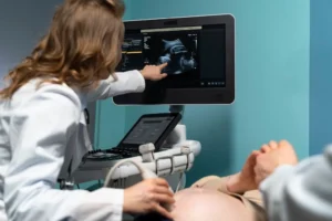
USG
Ultrasound Sonography, also known as an ultrasound scan, is a non-invasive imaging technique using high-frequency sound waves to generate images of internal body structures thus aiding in diagnosis and monitoring various conditions.
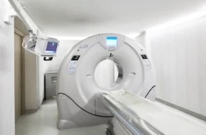
CT SCAN
Computed tomography scan, is an imaging technique that combines X-rays and computer technology to create detailed cross-sectional images (called slices) of the body. These images help visualize internal organs, bones, soft tissues, and blood vessels, thereby aiding in the diagnosis and treatment of various medical conditions.
DOPPLER
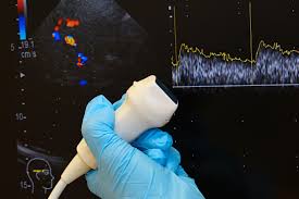
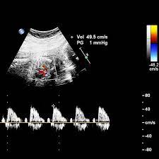
Doppler ultrasound is a medical imaging technique that deploys sound waves to visualize blood flow, using the Dopplers theory, which allows the assessment of blood flow in various parts of the body, including arteries and veins, and identify potential problems like blockages or narrowed blood vessels. Thus, Doppler ultrasound can be used in situations, including checking blood flow in pregnancy, diagnosing deep vein thrombosis (DVT), and assessment of blood flow to organs.
Colour Doppler is a type of Doppler ultrasound that uses colour to represent blood flow direction and velocity, making it easier to visualize and interpret the observations.
FETAL ECHO
A fetal echocardiogram, is an imaging technique used to examine the heart of a the developing fetus. It covers the assessment of the heart chambers, valves, and major blood vessels. It is also used to evaluate the heart’s rhythm and function, thereby helps doctors identify and diagnose congenital heart defects much before birth.
This test is important when there’s a family history or a routine prenatal ultrasound results in concerns about the baby’s heart.
This test id generally conducted between 18 and 24 weeks of pregnancy.
2D ECHO
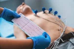
2D echocardiogram, is a non-invasive diagnostic test that uses sound waves to produce images of the heart. These sound waves are emitted from a transducer and bounce back from the heart creating a visual representation on a monitor. The images are displayed in two dimensions, showing the heart’s movement and help doctors to assess its various components.
2D echoes for several reasons:
- Diagnosing heart conditions:
Assessment of abnormalities like valve disorders, heart failure, congenital heart defects, and other structural problems.
- Monitoring heart health:
It can be used to monitor the progression of existing conditions of the heart or the effectiveness of treatments administered.
- Assessing heart function:
Evaluation of the hearts ability in pumping blood and identify any potential issues with blood flow.
SONOMAMMOGRAM
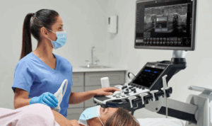
A sonomammogram, is non-invasive and radiation-free procedure of diagnostic imaging that uses high-frequency sound waves to create images of the breast tissue. It is used to supplement mammography, when breast tissue is dense. Sonomammography helps in the early detection and diagnosis of breast abnormalities.
OUR SPECIALISTS

Dr. Rajesh Murthy
FRCR(London)
Chairman
